Teratomas
Introduction
A teratoma is an encapsulated tumor with various tissue or organ components resembling normal derivatives of all three germ layers. The tissues of a teratoma may be quite different from surrounding tissues. Teratomas have been reported to contain hair, teeth, bone and, very rarely, more complex organs such as eyes, torso, and hands, feet, or other limbs.
Teratomas are usually present at birth (congenital), but small ones are often not discovered until much later in life.
Classification
Regardless of location in the body, a teratoma is classified according to a cancer staging system. This is helpful for treatment planning. Teratomas commonly are classified using the Gonzalez-Crussi grading system:
• 0 or mature (benign)
• 1 or immature, probably benign
• 2 or immature, possibly malignant (cancerous)
• 3 or frankly malignant.
Iranian specialists have made remarkable achievements in infants' surgeries and Iran is among few countries that have gained access to the latest surgical technologies
Teratomas are also classified by their content:
• A solid teratoma which contains only tissues
• A cystic teratoma which contains only pockets of fluid or semi-fluid such as cerebrospinal fluid, sebum, or fat
• A mixed teratoma contains both solid and cystic
Diagnosis
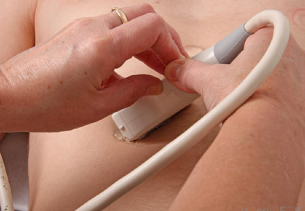
Although thought to be present since birth; but many teratomas are not diagnosed until much later in childhood or in adulthood. Sacrococcygeal and cervical teratomas are often detected by prenatal ultrasound. Additional diagnostic studies may include prenatal MRI.
Symptoms:
In the fetal life:
• The tumor is so large that the fetus may be damaged or dies.
• Large sacrococcygeal teratomas – in this condition, a significant portion of the fetus’ blood flow can be redirected toward the teratoma (a phenomenon called steal syndrome), causing heart failure, or hydrops, of the fetus. In some rare cases, fetal death can occur.
Beyond the newborn period:
• After newborn period, the symptoms of a teratoma depend on its location and organ of origin.
• Ovarian teratomas – present with abdominal or pelvic pain, caused by torsion of the ovary or irritation of its ligaments.
• Testicular teratomas – present as a palpable mass in the testis
• Mediastinal teratomas – can often cause compression of the lungs or the airways and may present with chest pain or respiratory symptoms.
• Some teratomas contain yolk sac elements, which secrete alpha-fetoprotein (AFP). Detection of AFP can confirm the diagnosis and is often used as a marker for recurrence or treatment efficacy.
Time of presentation
Teratomas of germ cell origin usually present in adult men and women, but they may also be found in children and infants. However, teratomas of embryonal origin are most often found in babies at birth and also in young children.
Complications
Teratomas are not usually dangerous for the fetus unless there is either a mass effect or a large amount of blood flow diverted to the tumor (known as vascular steal). The mass effect frequently consists of obstruction of normal passage of fluids from surrounding organs. The vascular steal can take toll on the growing heart of the fetus, even resulting in heart failure.
Even after surgery, there is a risk of regrowth in the same site, or in nearby organs.
Treatment
• Surgery
The treatment of choice is complete surgical resection of the tumor. Teratomas normally are well encapsulated and rarely invade the surrounding tissues, hence they are relatively easy to separate from surrounding tissues. Exceptions to this include teratomas in the brain, and complex teratomas that have become incorporated with adjacent muscles and other structures.
Prevention of recurrence does not require resection of surrounding tissues.
• Chemotherapy
For malignant teratomas, surgery is usually followed by chemotherapy.
Teratomas which are in surgically inaccessible locations, or are very complex to remove, or are likely to be malignant are sometimes treated first with chemotherapy.
Tumour markers:
Teratomas of germ cell origin usually present in adult men and women, but they may also be found in children and infants.
Depending on the tissue contents in the teratoma, the tumour may secrete a variety of chemicals having systemic effects. Some teratomas secrete the pregnancy hormone, called as human chorionic gonadotropin (beta-hCG). This can be used in clinical practice to monitor the successful treatment or relapse in patients with a known HCG-secreting teratoma. This hormone is not recommended as a diagnostic marker for all tumors, as most teratomas do not secrete it.
Some teratomas secrete thyroxine, and in some cases, this can lead to clinical hyperthyroidism in the patient.
In some cases, AFP can be used as a diagnostic marker specific for the presence of yolk sac cells within the teratoma. These cells can develop into a frankly malignant tumor known as yolk sac tumor or endodermal sinus tumor.
Adequate follow-up of teratoama includes close observation, repeated physical examination, scanning studies (ultrasound, MRI, or CT), and measurement of AFP or beta-hCG hormones.