Lymphangioma
Introduction
Lymphangioma refers to a rare, congenital condition that usually does not pose a health problem unless it occurs in a fetus (fetal cystic hygroma).
Lymphangiomas occur as a result of a blockage in the lymphatic system which causes fluid accumulation. They typically appear as a fluid-filled, yellowish or reddish bubble or mass beneath the skin.
This condition may affect people of all ages, but the majority of cases are diagnosed before age five. Lymphangiomas may affect any part of the body, but their most common site is on the neck or groin.
As they have no chance of becoming malignant, lymphangiomas are usually treated for cosmetic reasons only. However, when they occur early in the process of fetal development, cystic lymphangiomas can result in fetal demise due to excess fluid accumulation.
Iranian specialists have made remarkable achievements in infants' surgeries and Iran is among few countries that have gained access to the latest surgical technologies
Signs and symptoms
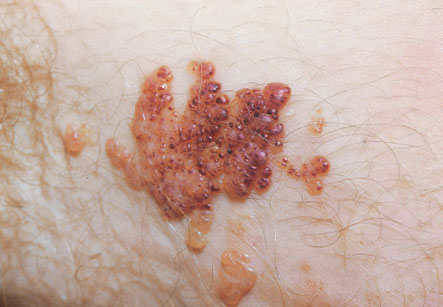
The lymphangioma comes in three distinct types, each of which has a slightly different appearance and different implications.
Lymphangioma circumscriptum consists of one or a group of translucent, bubble-like structures of varying sizes on the surface of the skin. They are usually pink to dark red in color. This type usually occurs on the skin surface while the other two types of lymphangiomas occur deeper in the skin.
Cavernous lymphanioma and cystic lymphangioma (also called cystic hygroma, familial nuchal bleb, fetal cystic hygroma, or hygromacolli) do not affect the skin surface like lymphangioma circumscriptum does. Instead they result in a bulge or thickening of the skin.
Cavernous lymphaniomasmay vary widely in size, ranging from a few centimeters in diameter to several centimeters wide. In some cases, they may even affect an entire extremity such as a hand or foot. They are usually painless, but the patient may feel mild pain when pressure is exerted on the area.
Cystic lymphangiomasis similar to cavernous lymphanioma. However, cystic lymphangiomas usually have a softer consistency than cavernous variety. This term is typically the one which is applied to lymphangiomas that develop in fetuses.
Causes and risk factors
The direct cause of lymphangioma is a blockage of the lymphatic system during development of fetus, although symptoms may not become visible until the birth of the baby. This blockage is thought to be caused by a number of factors, such as maternal alcohol use and viral infections during pregnancy.
Cases of cystic lymphangioma which emerges during the first two trimesters of pregnancy may be associated with genetic disorders including Turner?s syndrome, Noonan syndrome, and chromosome abnormalities such as trisomies 13, 18, and 21.
Prognosis
The prognosis for cases of lymphangioma circumscriptum and cavernous lymphangioma is excellent. There is a minor risk of infection, but this can be treated easily. The tumor can be removed at any time so long as there is no infection.
The prognosis is also good for fetuses that are diagnosed with cystic lymphangioma late in the pregnancy. However, this condition is dangerous when it develops early in a pregnancy, and is especially worrisome if there is an associated with genetic disorder. Most of the prenatal cases of cystic lymphangioma resolve on their own by about 18-20 weeks of gestation and their prognosis is moderate to good. But cases that are not resolved by this stage have a poor prognosis.
Diagnosis and treatment
The doctor can diagnose the cases of lymphangioma usually by inspection. In prenatal cases, cystic lymphangioma can be diagnosed using ultrasound technology. After detecting fetal cystic lymphangioma, amniocentesis or CVS procedures may be performed to check for associated genetic disorders. Draining fluid inside the lymphangiomas provides only temporary relief; therefore surgical resection is required to correct this condition.
Additional testing during pregnancy in cases of cystic lymphangioma:
Serial ultrasounds are needed to follow the baby’s growth, monitor the size of the tumour, and detect other congenital defects. A fetal MRI is recommended to differentiate between a cystic hygroma and cervical teratoma and also to assess the airway.
Special considerations during the delivery of babies with this condition:
Airway management at birth is critical in presence of giant neck masses. It is recommended that the delivery should be planned at a hospital equipped for high-risk deliveries as well as having pediatric and fetal surgical subspecialties.
The prognosis is also good for fetuses that are diagnosed with cystic lymphangioma late in the pregnancy.
Giant neck masses may compress the esophagus of fetus which causes polyhydramnios and may cause preterm labor. Giant cervical lymphangiomas are evaluated by ultrasound and fetal MRI to assess the possibility of airway compromise at birth. If there is concern about establishing an airway, an EXIT Procedure (ex-uterine intrapartum) is recommended. If an endotracheal tube cannot be inserted, a fetal tracheostomy needs to be performed. Occasionally, partial resection of the mass on continued placental support is required to place a tracheostomy.