Atypical Teratoid/ Rhabdoid Tumor ATRT
Introduction
Atypical teratoid/ rhabdoid tumor or ATRT is a very rare, fast-growing tumor of the brain and spinal cord. Although ATRT can occur in older children and adults, it is most common in children under 2 years of age.
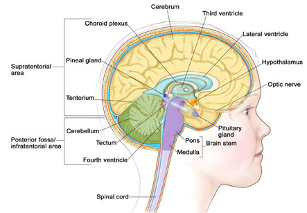
The preferred site of the tumor in the brain is cerebellum or brain stem. The cerebellum is the part of the brain concerned with movement, balance, and posture. The brain stem controls breathing, heart rate, and the nerves and muscles used in seeing, hearing, walking, talking, and eating. The other parts of the central nervous system (brain and spinal cord) can also be involved in this tumor.
Causes:
Atypical teratoid/ rhabdoid tumor may be linked to a change in a tumor suppressor gene called SMARCB1. This gene makes a protein that helps to control cell growth. Mutation or changes in the DNA of tumor suppressor genes leads to this cancer.
These genetic changes in the SMARCB1 gene may be inherited. In case of inheritance, the tumor may form in two parts of the body at the same time.
Iranian specialists have made remarkable achievements in infants' surgeries and Iran is among few countries that have gained access to the latest surgical technologies

Symptoms of atypical teratoid/ rhabdoid tumor:
The atypical teratoid/ rhabdoid tumor is fast growing tumor, therefore the symptoms develop quickly and progress to severity over a period of days or weeks. Usually these are:
1. Morning headache or headache relieved after vomiting.
2. Nausea and vomiting.
3. Unusual sleepiness.
4. Increased head size in infants
5. Loss of balance, lack of coordination, and problems during walking.
Diagnosis:
The following tests and procedures may be used:
1. Physical examination and history: A thorough clinical examination of the patient is done to evaluate general health. A history of the patient’s health habits and past illnesses and treatments is also asked.
2. Neurological exam: this is important for checking the brain, spinal cord, and nerve function. Also, the mental status of the patient, coordination, and gait, as well as the muscle power, sensations and reflexes are checked.
3. CT scan: In this procedure, a series of detailed pictures of areas inside the body are taken by a computer linked to an x-ray machine. A dye may be injected into a vein or swallowed to show the organs or tissues more clearly.
4. MRI (magnetic resonance imaging): This procedure uses a magnetic energy and a computer to make a series of detailed pictures of areas inside the brain and spinal cord. MRI is a preferred imaging study for brain and spinal cord tumors.
5. Lumbar puncture: This procedure is used to collect the cerebrospinal fluid from the spinal column by injecting a hollow needle into the spinal column. It can give information about brain metabolism and infections.
6. Ultrasound exam: High-energy sound waves are bounced off internal tissues or organs and echoes made by various soft tissues are represented in the form of sonogram. A renal ultrasound is useful to check whether the AT/RT has developed in the kidneys at the same time as that in the brain.
7. INI1 gene testing: A laboratory test in which a sample of blood or tissue is tested for the INI1 gene.
The prognosis or chances of recovery depend on the following factors:
1. Age of the child.
2. The extent of tumor remaining after surgery.
3. Spread of the cancer to other parts of the brain and spinal cord or to the kidney.
Treatment of the Atypical teratoid/ rhabdoid tumor:
There are four treatment modalities for this cancer as follows:
1. Surgery- Neurosurgery is used to diagnose and treat atypical teratoid/ rhabdoid tumor. The surgeon removes the entire visible tumor at the time of the surgery. However, most patients are given chemotherapy and may be radiation therapy after the surgery to kill any cancer cells left behind. This treatment given after the surgery, to prevent cancer recurrence is known as adjuvant therapy.
2. Chemotherapy- This treatment uses drugs to kill the cancer cells or stopping their multiplication. This therapy can be taken orally or intravenously. Chemotherapy can be given directly into the cerebrospinal fluid, to kill the cancer cells in the brain and spinal cord. This is called as intra-thecal chemotherapy.
3. Radiation therapy- This is the treatment using high-energy x-rays or other types of radiation to kill cancer cells or prevent them from growing. However, radiation therapy can affect growth and development of brain in young children, the dose of radiation therapy is lower than usual.
4. High-dose chemotherapy with stem cell transplant- This can be used to first kill the tumor cells by high dose of drugs and replacing the normal blood forming cells killed by chemotherapy. The stem cells are taken from the bone marrow of healthy donor.
5. Genetic counseling- This therapy is recommended for ATRT. A discussion with a trained professional about inherited diseases is done and a possible need for genetic testing is determined.