Knee Arthroscopy
Introduction
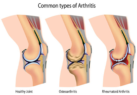
Knee arthroscopy is a procedure to diagnose the pathology or correct certain conditions in arthritic, painfully injured or stiff knees. The knee joint is composed of the lower end of the thigh bone or femur, the upper end of the shin bone or tibia and the knee cap or the patella. There is a complex system of cartilage, ligaments, tendons or fibrous attachments and muscles that keep the knee joint steady and mobile. The cartilage between the joint also known as the menisci and the ligaments called the cruciate ligaments can be damaged due to injury.
A knee arthroscopy can be done to repair or replace this cartilage or repair the ligament. Arthroscopy can also be done to realign a misplaced or dislocated knee cap, remove growths or Baker’s cysts and repair small fractures. Knee arthroscopy is commonly done to assess the pathology in chronically painful and stiff knees.
Iran ranks among the top 10 countries in orthopedics and Iranian surgeons perform high quality orthopedic surgeries at highly affordable prices
Procedure
This is a short operation usually performed in Day care Units. Patient may need someone to drive them home after the operation. Usually local anesthesia is used where the knee is numbed with an injection of drugs and patient may be given anti anxiety medicines to relax while the operation is performed painlessly. Some centers may also use spinal anesthesia where the anesthetic drug is injected between the vertebrae and it makes them numb below the waist. In rare cases general anesthesia may be used wherein they are rendered unconscious during the operation.
Under the guidance of the camera the repair of the ligaments and cartilages are undertaken.
A tourniquet or cuff like device will be tied around the thigh to make the operative site blood-less and minimize blood loss. Two or three small incisions are made over the knee and saltwater or saline will be pushed in through these openings to open up the joint for better viewing. A narrow arthroscope or tube with a camera attached on its tip will be inserted through one of the openings. The tube is connected to a video monitor in the operation theatre by which surgeon can be guided through the procedure. If the patient wishes they can also see the procedure on this monitor.
In case of a diagnostic procedure the surgeon can view the problem in the joint directly. If this is a corrective procedure the other small incisions are used to insert operative instruments. Under the guidance of the camera the repair of the ligaments and cartilages are undertaken. At the end of the surgery the saline will be drained out, the incisions sutured and the wounds dressed.
Recuperation and post operative complications
Most people can be discharged on the same day of the surgery. Patient is advised to avoid heavy activities or exercise during the first few weeks after surgery. They may be given pain relieving medicines for the pain and you can also apply ice packs and elevate the leg to ease the pain and swelling.
Risks associated with most surgical procedures are also present with a knee arthroscopy operation. This includes bleeding, pain, blood clots and nerve damage. The benefits of this procedure include the fact that it is less invasive than an open repair or correction of meniscal tears, ligament damage or dislocated knee cap. Recovery and return to normality is rapid compared to an open surgery. A diagnosis made on the basis of an arthroscopy can often be more accurate than with a simple X ray.
For diagnosis of ailments of the knee an Ultrasonography (USG), CT scan or Magnetic resonance Imaging (MRI) can be cited as alternatives. With the use of contrast dyes these procedures can be made more accurate in diagnosis. Use of braces, pain killer medicines, physiotherapy and warm compress can be alternatives to knee injuries that lead to torn ligaments or menisci.