Vitreo Retinal Surgery
Introduction
Conditions commonly evaluated and treated by retinal specialists or vitreo-retinal surgeons usually include retinal tears and detachments, diabetic retinopathy, macular degeneration, ocular trauma, ocular tumors, inflammatory ocular conditions, hereditary retinal diseases and a variety of less common, highly complex disorders. Today, highly advanced, state-of-the-art techniques are available to treat these vision threatening diseases. Vitreoretinal Surgery deals with diseases involving the vitreous humour and the retina.
The retina has two parts: the peripheral retina and macula. The macula is a spot on the retina almost near its centre. The macula is very small. The large area of the retina that surrounds this spot and makes up 95% of the retina is called as peripheral retina.
The peripheral retina gives us vision to the side (peripheral vision). This part of the retina is at work when we see something out of the corner of our eye. However, the peripheral retina is not able to see minor details clearly. Therefore, the peripheral field of vision cannot be utilized to read, thread a needle, drive, or even recognize a face. If you see someone out of the corner of your eye, you might be able to tell who it is as the general shape of the person can be recognized, but the expressions on that person’s face cannot be seen.
Iran is now ranking first both in the region and in the Middle East and the Iranian ophthalmology science is among the world top majors
The macula is responsible for central vision. The visual acuity is greatest at the macula. The details of object, its color, brightness as well as depth are best perceived at macula.
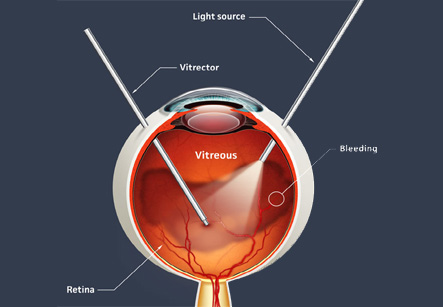
The vitreous is much like the clear ‘white’ of an egg. It fills the central cavity of the eye. The vitreous is attached to the retina. It is most strongly attached in the back part of the eye to the optic nerve, the macula and the large retinal blood vessels.
Need of retinal or vitreous surgery
Retinal and vitreous surgery addresses problems of eye such as Retinal Detachment and deep Intraocular Infection. Retinal and vitreous problems can cause severe loss of vision or even blindness. In some cases, surgery may be beneficial and it may also prevent severe loss of vision. However, for preserving vision, the surgery needs to be performed at early stages of retinal disease. Most serious retinal problems that require surgery are caused by problems with the vitreous.
Patients who have had cataract surgery have about a 1 – 2% chance of developing retinal detachment.
When there is a tear of the retina, the liquid in the vitreous cavity may pass through the tear and get accumulated under the retina. The liquid collects under the retina and lifts it off from the posterior wall of the eye. Gradually, the liquid from the vitreous passes through the retinal tear and settles under the retina, separating it from the back wall of the eye. This separation of the retina is called as retinal detachment.
Vision is lost wherever the retina becomes detached. Most of the retinal tears are located in the peripheral retina. Therefore, the retinal detachment first results in loss of side (peripheral) vision. A patient may notice a dark shadow or a veil coming from one side, above or below. In most cases, the entire retina will eventually detach and all useful vision in the affected eye will be lost.
Certain people have a greater chance of getting a retinal detachment than others. These patients are those with:
1. A high degree of nearsightedness (myopia)
2. Family history of retinal detachment
3. Retinal detachment in the other eye
4. Thinning of the retina due to lattice degeneration or other degenerative disorders of the retina
A person in any of these high-risk groups should have regular retinal examinations. These people should see the eye care specialist immediately if they experience even slight loss of peripheral vision, or see sudden flashing of lights or spots.
When an infection occurs inside the eye, it is called ‘endophthalmitis.’ Endophthalmitis often occurs after intraocular surgery or penetrating trauma.
Endophthalmitis can be treated with antibiotics. These antibiotics can be administered in a variety of ways, from eyedrops to injections. In severe non-responding cases, a vitrectomy is performed to remove the infectious material from inside the eye. The doctor will discuss the best method of treatment with the patient.