Medical Retina
Introduction
The retina is situated at the back wall of the eyeballs which receives light rays from the front of the eye and sends them through the optic nerve to the brain. In brain, they are turned into clear, bright, colorful images. The vitreous is a clear, jelly-like substance that lies between the eye’s natural lens and the retina. When disease or injury causes clouding of the vitreous or damage to the retina, it can cause partial or complete loss of vision.
In case if the primary eye doctor finds signs of a retinal disorder during a general eye exam, the patient is usually referred to a retina specialist for additional tests and possible treatment. Early detection and treatment is essential for treating retinal disorders as once the retina is damaged, repair is extremely difficult.
Preparation for the Examination
A visit to the retina specialist starts with the creation of a patient record. This includes patient’s address and telephone numbers, names of other doctors, list of medications being taken, and past medical records if available. Some doctors even ask the patient to bring all the medications currently being used. It is prudent to bring sunglasses to wear as the retina exam includes dilating the eyes.
Iran is now ranking first both in the region and in the Middle East and the Iranian ophthalmology science is among the world top majors
The Examination
A retina exam is focused on the back of the eye. The doctor spends a lot of time looking through the pupil at the vitreous, retina and other structures located in the back portion of the eye. The eyes are dilated with medications so that the doctor can get a clear view of these structures.
The following methods are normally used in diagnosing retina problems:
1. Indirect ophthalmoscopy- it uses a specialized microscope which allows the doctor to observe the vitreous, retina and other internal structures of the eye. The ophthalmoscope is usually the first instrument used by the doctor during the exam because it provides an overview of the situation.
2. A visual field or perimetry test- this test measures the ability of the eye to see straight ahead and to the side (peripheral vision). During a visual field test, one eye is temporarily covered and the patient is asked to look straight ahead at a fixed space. There are types of this test:
• The moving target test- the targets are moved from the side, where they are not visible, toward the center of vision until the patient sees them. This test can be performed with the help of either a black screen on the wall or with a large bowl-shaped instrument.
• A fixed target test (computerized static perimetry)-During this test, the patient sits in a chair facing a computer screen. This test uses small points of light that appear bright or dim but do not move.
The visual field examination is useful to detect blind spots that may be caused by diseases of the retina.
1. Fluorescein angiography – this test is used to examine blood vessels in the retina. A fluorescein dye is injected into the blood stream and as the blood circulates through the retina, a series of rapid, sequential photographs of the eye are taken. This test provides useful anatomic and functional information about the retina.
2. B-scan ultrasound- this is a method for viewing the structures at the back of the eye making use of high frequency sound waves. This technology provides a topographical view of the eye and also the information which is difficult to achieve.
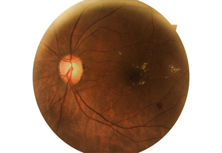
3. Fundus photography- this method uses specialized film and digital cameras for diagnosing disorders of retina. It is important in monitoring the progress of certain retinal diseases and also to evaluate the treatments started.
Upon completion of tests required for the diagnosis, the doctor discusses the various treatment options available for the disorder found.
All tests need not be done in each patient. The choice of tests depends on medical history, suspected diagnosis, and also the preference of the specialist.
Upon completion of tests required for the diagnosis, the doctor discusses the various treatment options available for the disorder found. Although retinal disorders are often complicated to treat, modern medicine has made advanced treatments available in recent years.
Common Retina Disorders diagnosed by these methods are diabetic retinopathy, age-related macular degeneration, retinal detachments, glaucoma, and some congenital disorders.