Cataract Surgery
Introduction
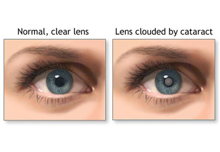
In a normal eye there is a clear lens that allows light to pass and aids in vision. When this glassy lens becomes clouded due to age, injury, drugs or some other causes the condition is called cataract. Cataract surgery essentially replaces the natural lens that is afflicted with cloudiness with an artificial lens shaped similarly.
Surgery Procedure
The surgery takes place under local anesthesia wherein the muscles and tissues around the operated eye is numbed. The eyelids are kept open using an instrument called the speculum. BSS or Balanced Salt Solution is used to keep the eye surface moist during the operation. The eye is placed under a microscope. The surgeon operates under the microscope.
There are two common types of cataract surgery. One of the types is called “extracapsular” type of cataract surgery. Here the surgeon makes a small slit on the side of the cornea. Cornea is the transparent layer or membrane that lies over the lens.
Iran is now ranking first both in the region and in the Middle East and the Iranian ophthalmology science is among the world top majors
Usually both eyes are not operated at one go. The eyes are operated eight weeks apart.
Thereafter using instruments the affected lens is slipped out of the slit and an artificial lens called IOL or Intraocular Lens is inserted in its place and secured. The slit is then sewn up. The IOL is made up of plastic, silicone or acrylic. This lens can stay on in the eye for life.
The second type that is one of the commonest these days is called “phacoemulsification”. It needs a smaller incision than the first type. In this type the clouded lens is first liquefied using certain instruments and then the liquefied mass is sucked out of the small slit. The instrument used send out ultrasound vibrations to the lens to break it into small pieces and liquefy it. These vibrations are too tiny to be felt.
Recuperation and post operative complications
The surgery usually is short and patient may be discharged on the day of the operation. After the operation eye drops are applied and the eye is dressed. Usually both eyes are not operated at one go. The eyes are operated eight weeks apart.
After the surgery there may be some dryness and itching in the eyes. In order to heal effectively some medications that keep the pupil dilated are applied as drops to the operated eye. These drops may also lead to discomfort due to too much light exposure into the eye. Patients are given dark glasses to wear after the surgery. This protects their eyes from exposure to too much light.
Pain can be relieved by pain relieving medication. Antibiotics are prescribed orally as well as in the form of eye drops and ointments to prevent infection after the surgery. Patient is advised to avoid heavy physical work and exercise during the initial weeks after surgery.
Around a month after the surgery the maximum vision possible with the new lens is achieved. Most patients may need glasses to achieve further correction of vision.
In some patients the new lens may get clouded over time. The operation may be re-done for these patients. Usually the lens remains good and requires no care for the rest of the life.