Cerebrovascular Pathology
Introduction
Cerebrovascular disease is the 3rd most common cause of death all over the world. It is most common of all CNS diseases considered together.
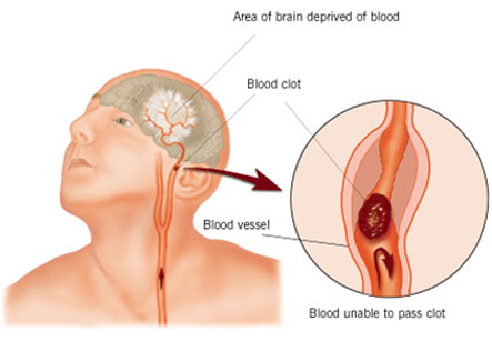
Stroke is a sudden and dramatic development of a focal neurological deficit as a result of vascular impairment. It is estimated that the stroke is responsible for 10% of all deaths occurring due to organic causes. The main causes of stroke include hypertension, cerebral atherosclerosis, or both.
Symptoms of stroke may include the following:
1. Sudden weakness or numbness of the face, arm, or leg on one side of the body
2. Loss of speech, or difficulty in speaking or understanding speech
Iran has made significant progress in neurosurgery and The world renowned Iranian scientist in neurological surgery Professor Majid Samii has garnered the 2014 Golden Neuron Award. We have all the information you need about public and private clinics and hospitals that provide Neurosurgical surgeries in Iran, Islamic Republic Of with the best quality and lowest possible price
3. Dimness or loss of vision, in one of the eyes
4. Unexplained dizziness
TIAs or “transient ischemic attacks” is a name given to reversible focal neurological symptoms secondary to ischemia which may last from several seconds to 24 hrs.
Arterial infarction:
Main causes of arterial infarction include thrombosis and embolism.
Factors contributing to infarction include:
1. Decreased oxygen in blood
2. Reduced hemoglobin in blood
3. Hypoxemia
4. Decreased perfusion as a result of cardiac arrest, hypotension, shock, pulmonary embolism, CHF and raised ICP
5. Altered composition of blood due to polycythemia, thrombotic thrombocytopenic purpura, macroglobulinemia, treatment with oral contraceptives, pregnancy, and puerperium
6. Misc. causes of infarction
7. Hypoglycemia
8. Vasculitis
9. Endarteritis obliterans
10. Compression of vessels by tumors or aneurysm
Major sources of emboli
1. Atria, cardiac valves, and mural emboli
2. Aorta and Neck vessels
3. Peripheral vessels – which are rare
Size and extent of infarcts is influenced by:
1. Site of the occlusion
2. Presence and efficacy of collateral circulation
3. Rapidity of occlusion
There are two main types of infarcts:
Infarcts of end-arterial zones
Infarcts at boundary zones (water-shed) infarcts
Classification of Infarcts
According to type
1. Anemic (Pale)
2. Hemorrhagic (red)
According to site of involvement in brain
1. Cortical and subcortical
2. Cortical only
3. White matter only – HTN etiology
4. Basal Ganglia
Main stages in the evolution of cerebral infarcts are:
Stage I: Coagulative necrosis with edema
Ischemic cell change
1. Nuclear pyknosis with cytoplasmic eosinophilia – eosinophilic neurons
2. Pallor of nuclear and cytoplasmic nucleic acids – ghost neurons
3. Shrinkage and condensation of the perikaryon – dark neurons
4. Precipitation of formaldehyde pigment on the neuronal perikaryon
5. Neuronal vacuolation
6. Development of a perineuronal halo
Stage II: Liquefaction and phagocytosis
Stage III: Cavitation and shrinkage
1. Cavitation probably results as the astrocytes cannot grow on a dead substrate
2. Various components of the brain react in different ways to the same ischemic injury
Cerebral and meningeal hemorrhage
3 main non-traumatic causes of haemorrhage include hypertension, vascular malformation and blood dyscrasias
Hypertension can cause vascular changes and it is the most important etiological factor for infarction and haemorrhage. Hypertension accentuates atherosclerosis of the large arteries.
Chronic hypertension also alters the wall of the arterioles in the following ways:
1. Hyaline degeneration
2. Fibrinoid necrosis
3. Charcot-Bouchard aneurysm – this type of aneurysm is most frequently found in the small arteries of the basal ganglia
4. Thrombotic obliteration of altered vessels
Sites of hypertensive hemorrhage may include:
1. Putamen
2. Thalamus
3. Cerebral hemispheric white matter
4. Pons
5. Cerebellar white matter
The location and relative incidence of hypertensive hemorrhages is in accordance with the distribution of Charcot-Bouchard aneurysms in brain.
Complications of hypertension may include rupture of vessels into ventricles or through cortex into leptomeninges.