Cardiotomy
Introduction
A cardiotomy is a cut into the heart to access it for surgical procedures or provide a route for suction. This procedure is necessary in a variety of cardiovascular surgeries to deal with conditions of the heart and neighbouring major blood vessels. Surgeons perform this incision with care so it will be easier to suture it at the end of the procedure, and also to reduce the risks of post-surgical complications. Advanced knowledge of heart anatomy and considerable experience in cardiovascular surgery is required for making an incision into the cardiac wall, as proper placement of the cut is critical.
Some surgeries may require a cardiotomy so that the surgeon can access the necessary anatomical structures. This may be required for repair of a birth defect or acquired problem with the heart. Surgeons make incisions as small as possible to protect the patient. The focus of surgery is on careful working inside the limited space available. Once the procedure is complete and the repair appears to be satisfactory, the site can be sutured. If the cut is in too deeper plane or placed at the wrong angle, it may prove risky to the patient. The surgeon plans before the surgery to make sure that it is performed appropriately.
Iran is among the top 10 countries in treating cardiovascular diseases, while it ranks first in the Middle East

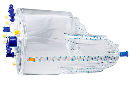
Another use for a cardiotomy is to create an adequate area for suction. Cardiovascular surgeries are notorious as they can cause flows of blood in and around the heart. This can obscure the surgical field and makes it hard to work. With suction equipment, surgical assistants clear the field continuously so the surgeon can clearly see. It is important to have easy access for suction that won’t get in the surgeon’s way or endanger the patient.
Historically, blood collected at a cardiotomy site used to be filtered and recycled for replacing at least some of the patient’s lost blood. Many researchers believe that this might not be the best medical practice, as such replaced blood can introduce cellular debris, including even small chunks of lipids. This can lead to strokes and other problems afterwards. In addition, it appears to contribute to various coagulation defects which can create need for more transfused blood from a donor than it would otherwise be necessary. In some procedures or in some hospitals, routine recycling of cardiotomy blood is no longer recommended.
Like other measures taken during surgery, a cardiotomy is also not without risks. Patients may experience inflammation and infection that can result in severe pain, fever, and nausea. Poor placement of cardiotomy can damage the heart or neighboring structures. Rupture of the sutures can cause extensive bleeding and other complications. After surgery on the heart, it is necessary that the patient should stay in the hospital for a few days for monitoring. Some precautions need to be observed upon being discharged to prevent complications and identify them early if they develop.
Cardiotomy syndrome:
The post cardiotomy syndrome is observed in patients about 4-6 weeks after undergoing cardiac surgery (especially repair of atrial septal defects). There may be pericarditis and a pericardial effusion which may result in cardiac tamponade. This condition is usually suspected in any child who presents with flu-like syndrome 4-6 weeks after cardiac surgery. The syndrome is closely related to Dressler’s syndrome.
Postpericardiotomy syndrome (PPS):
The post cardiotomy syndrome is observed in patients about 4-6 weeks after undergoing cardiac surgery (especially repair of atrial septal defects).
This is a febrile illness secondary to an inflammatory reaction which is set in the pleura and pericardium. It is more common in patients who have undergone cardiac surgery which involves opening the pericardium. However, this post-pericardiotomy syndrome has also been described following myocardial infarction (called as Dressler syndrome). This can also occur as a rare complication after percutaneous procedures such as coronary stent placement, implantation of epicardial pacemaker leads and transvenous pacemaker leads, following blunt injuries, stab wounds, and heart puncture wounds.
Pericardial effusions usually accompany this syndrome and may develop into early or late post-operative cardiac tamponade and even recurrent cardiac tamponade. The syndrome is also characterized by pericardial or pleuritic chest pain, friction rubs on auscultation, pleural effusions, pneumonitis, and abnormal ECG as well as radiography findings.