Cardiotocography
Introduction
Cardiotocography is the technique used to monitor a baby’s heart rate and a mother’s contractions while the baby is in the uterus. Cardiotocography, also known as CTG, uses ultrasound technology to measure the baby’s heart rate. At the same time it also measures the contractions of the uterus. CTG can be used both antenatally (before birth) and during labour to monitor the baby for any signs of distress. By looking at various different aspects of the baby’s heart rate, doctors and midwives can monitor condition of the baby.
Procedure:
CTG is most commonly carried out externally. This means that the equipment used to monitor baby’s heart is placed on the abdomen of the mother. An elastic belt is placed around the mother’s abdomen. This belt has two round plates about the size of a tennis ball which make contact with the skin. One of these plates uses ultrasound to measure the baby’s heart rate. The other plate measures the pressure in the abdomen and the uterine contractions. The doctor or midwife puts some jelly between the plate and the abdomen to help get a strong signal.
Iran is among the top 10 countries in treating cardiovascular diseases, while it ranks first in the Middle East
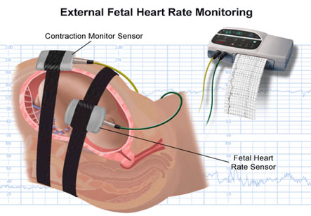
The CTG belt is in turn connected to a machine which interprets the signals coming from the plates. The baby’s heart rate can be heard as an amplified beating or pulsing sound which the machine produces. Some mothers in labour can find this sound distracting or worrying, but it is possible to turn the volume down if the noise disturbs the mother. The machine also provides a printout which shows the baby’s heart rate over a certain length of time. It also shows changes in the heart rate changes with the contractions. In case of CTG before the woman is in labour, the patient is asked to press a button on the machine every time the baby moves. At this time, there are no contractions of the uterus and the CTG will only monitor the baby’s heart rate.
CTG monitors the baby’s resting heartbeat and also its response to uterine contractions.
Sometimes, the fetal heart monitoring is very essential, but the signals cannot be found using the external monitor; internal monitoring can be used. For internal monitoring, a small device called as an electrode is inserted through the vagina and placed on the baby’s scalp. This device records the heart rate.
A thin tube called as catheter may also be inserted through the vagina into the uterus. This tube measures the strength of contractions. There may be some discomfort while the catheter is being inserted but it should not be too painful.
Mechanism of working of CTG:
Cardiotocography uses ultrasound energy to detect the baby’s heart rate. Ultrasound is a high-frequency sound that cannot be heard, but it can be emitted and detected by special machines.
Ultrasound travels freely through fluid and soft tissues. However, when the ultrasound waves hit a more solid surface or structure, they are reflected back (or bounce back as ‘echoes’). For example, the ultrasound will travel freely through blood in a heart chamber. But, when it hits a solid valve, a lot of the ultrasound echoes back.
As ultrasound ‘hits’ different structures in the body, of different density, it sends back echoes of varying strength. These echoes are detected by machine.
In CTG monitoring a special type of ultrasound, known as Doppler, is used. This type of ultrasound can be used to measure echoes from structures that are moving, making it useful for monitoring heart rate.
The other plate on the CTG measures how tense is the surface of the mother’s abdomen is. This measurement is used to show when the uterus is contracting.
CTG monitors the baby’s resting heartbeat and also its response to uterine contractions. It can be used in cases of:
• High blood pressure in the mother.
• The woman is expecting twins.
• The baby has passed meconium or stools into the amniotic fluid.
• The heart rate of the baby is suspected to be dropping with contractions.
• When epidural anesthesia is used for pain relief.
It is normal for a baby’s heart rate to vary between 110 and 160 beats a minute. This is much faster than the adult heart rate, which is about 60-100 beats per minute. A heart rate in the baby that doesn’t vary or is too low or too high may indicate a problem.
Changes in the baby’s heart rate occurring along with contractions form a pattern. Certain pattern of heart rate may suggest a problem, which is known as fetal distress. If test results suggest that there is distress, the doctor may decide to deliver the baby right away. This may mean that a caesarean section or assisted delivery using vacuum or forceps may be required for safe delivery of the baby.