Cardiac PET
Introduction
A heart positron emission tomography (PET) scan is an imaging study which uses a radioactive substance called as tracer to detect cardiac disease or poor blood flow in the heart. Unlike magnetic resonance imaging (MRI) and computed tomography (CT), which reveal the structure of and blood flow to and from organs, a PET scan shows how organs and tissues are functioning.
A heart PET scan can find out whether all the areas of the heart muscle are receiving enough blood and also if there is any heart damage or scar tissue in the heart.
Applications of cardiac PET scan:
• Cardiac Disease Diagnosis
Initial diagnosis of coronary artery disease is the primary reason for a cardiac PET scan study. This disease is characterized by blockages in the blood vessels that supply blood to the heart. The cardiac PET scan is extremely accurate in measuring the blood flow through the coronary arteries.
Iran is among the top 10 countries in treating cardiovascular diseases, while it ranks first in the Middle East
• Cardiac Disease Staging
The coronary artery disease must be staged after diagnosis in order to treat it properly. A cardiac PET scan can identify the extent and severity of coronary artery disease in the affected area. This helps in treatment planning.
• Cardiac Disease Follow-Up
A cardiac PET scan is also performed to evaluate the effectiveness of cardiac procedures such as coronary bypass graft surgery or coronary angioplasty. The bypass surgery replaces a clogged, diseased coronary artery by a healthy blood vessel from other part of the body. The angioplasty opens a clogged, diseased artery by a balloon which is inserted and inflated directly in the blood vessel. A PET scan can ensure adequate blood flow to the heart after bypass surgery or angioplasty.
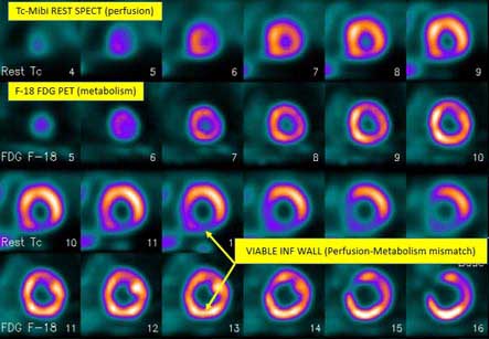
• Myocardial Viability
A cardiac PET scan is commonly performed to assess myocardial viability of patients who have suffered cardiac damage from a heart attack. Data from this test may determine whether the patient will benefit from an angioplasty, bypass surgery, or only medical treatment.
Preparation:
The patient is instructed not to eat anything for 4 – 6 hours before the scan. You can drink water if needed.
The doctor should be informed if:
1. The patient is afraid of close spaces (having claustrophobia). In such cases, sedatives may be given to help the patient feel sleepy and less anxious.
2. If the patient is pregnant or might be pregnant.
3. There is allergy to injected contrast dye
4. If patient is on insulin for diabetes. Insulin and elevated blood sugar levels can give false results of PET scan.
5. Taking any medications, even without prescription. Sometimes, medications may interfere with the test results.
Procedure:
1. A PET scan requires a small amount of radioactive material (tracer). This tracer is given to the patient through a vein (IV), usually on the inside of the elbow. It travels through the blood and distributed in various organs and tissues, including the heart. The tracer helps the radiologist to see certain areas or diseases more clearly.
2. The patient needs to wait in the hospital as the tracer is being absorbed by the body. This usually takes about 1 hour.
3. Then, the patient is made to lie on a narrow table, which slides into a large tunnel-shaped scanner. Electrodes for an electrocardiogram (ECG) are placed on the chest. The PET scanner detects signals coming from the tracer. A computer converts the results into 3-D pictures. The images are displayed on a monitor for the doctor to read. The results can be printed or cut into a CD-ROM.
4. The patient must lie still during the PET scan so that the machine can produce clear images of the heart. The test takes about 90 minutes to complete.
Side effects:
A sharp sting may be felt when the needle containing the tracer is placed into the vein.
A PET scan causes no pain. Patient may feel uncomfortable on hard and cold table, but a blanket or pillow can be made available. An intercom in the room can allow you to speak to scanner operator at any time.
Risks:
The amount of radiation used in a PET scan is low. It is about the same amount of radiation as in most CT scans. Also, the effect of radiation does not last for very long in the body.
A cardiac PET scan is commonly performed to assess myocardial viability of patients who have suffered cardiac damage from a heart attack.
Women who are pregnant or are breastfeeding should inform this to their doctor before having this test. Infants and babies developing in the womb are more sensitive to the effects of radiation as their organs are still in growing phase.
Rarely, it is possible to have an allergic reaction to the radioactive substance. Some people may develop pain, redness, or swelling at the injection site.