Pelvic Ultrasound
Introduction
A pelvic ultrasound is an investigation which uses sound waves to visualize the organs and structures in the lower belly (pelvis).A pelvic ultrasound helps to look at the bladder, ovaries, uterus, cervix, and fallopian tubes of a woman.
Organs and structures which are solid and uniform (such as the uterusor ovaries) or that are fluid-filled (such as the bladder) show up clearly on a pelvic ultrasound. However, bones or air-filled organs, such as the intestines, do not show up well on an ultrasound and may also keep other organs from being seen clearly.
Pelvic ultrasound in females can be done two ways: transabdominal and transvaginal.
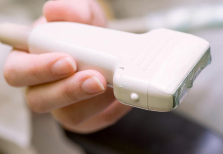
1. Transabdominal ultrasound. A small handheld device called as transducer is passed back and forth over the lower belly for diagnosing large uterine fibroids or other problems.
2. Trans-vaginal ultrasound. The transducer is shaped to fit into a woman’s vagina. Sometimes, both transabdominal and transvaginal ultrasounds are performed to look at the whole pelvic area. A transvaginal ultrasound is usually done to look for problems with fertility. In rare cases, a hysterosonogram is done to look at the inside of the uterus by filling the uterus with fluid during a transvaginal ultrasound. Sometimes, a small sample of tissue (or biopsy) is taken with small tools inserted through the vagina during this procedure.
We have all the information you need about public and private clinics and hospitals that provide gynaecological treatment in Iran
In both the types of pelvic ultrasound, the transducer sends the reflected sound waves to a computer, which converts them into a picture that is shown on a video monitor. These pictures or videos may be saved as a permanent record.
Indications:
For men and women, pelvic ultrasound may be done to:
1. Find the cause of blood in the urine (hematuria). An ultrasound of the kidneys may also be done
2. Find the cause of urinary symptoms
3. Look at the size of the bladder before and after urination. This can determine whether the bladder is emptying completely during urination
4. Check for growths in the pelvis
5. Evaluate colorectal cancer and how it is responding to treatment.
For women, pelvic ultrasound may be done to:
Find out the cause of pelvic pain
1. Look for the causes of vaginal bleeding
2. Look for evidence of pelvic inflammatory disease (PID)
3. Find a lost intrauterine device (IUD)
4. Determine the size and shape of the uterus and the thickness of the uterine lining (endometrium)
5. Check the size and shape of the ovaries
6. Check the condition and size of the ovaries during treatment for infertility
7. Confirm a pregnancy and its location. Pelvic ultrasound may be used early in pregnancy to check the gestational age or to diagnose a tubal pregnancy (ectopic pregnancy) or multiple pregnancy
8. Check the cervical length in a pregnant woman at risk for preterm labor
9. Check a lump found during a pelvic examination
10. Check uterine fibroids found during a pelvic examination
11. Guide a procedure to remove an ovarian follicle for in vitro fertilization
Before the procedure
• Notify the doctor if you are sensitive to or are allergic to latex.
• Generally, no fasting or sedation is required for a pelvic ultrasound, unless the ultrasound is used as a part of another procedure that requires anesthesia.
• For a transabdominal ultrasound, patient is asked to drink several glasses of water before the procedure. Full bladder is necessary for this procedure.
• For a transvaginal ultrasound, the bladder should be empty right before the procedure.
• Specific preparation may be needed depending on specific disorder of the patient, if any.
During the procedure
A pelvic ultrasound may be performed in the doctor’s office, on an outpatient basis, or as part of the stay in a hospital. Procedures may vary depending on the condition and hospital’s practices.
Patient is asked to remove any clothing, jewelry, or other objects that may interfere with the scan. Patient may need to change into surgical gown.
Trans-abdominal ultrasound:
1. Patient lies on the back on an examination table.
2. A gel-like substance is applied to the abdomen.
3. The transducer is pressed against the skin and moved over the area being studied.
4. When blood flow is being assessed by a Doppler probe, a “whoosh, whoosh” sound may be heard.
5. Images of structures are displayed on the computer screen.
6. Once the procedure has been completed, the gel is removed.
For a transvaginal ultrasound:
1. Patient lies on an examination table, with the feet and legs supported as for a pelvic examination.
2. A long, thin transvaginal transducer, covered with a plastic or latex sheath is lubricated. The tip of the transducer is inserted into the vagina. This may be slightly uncomfortable.
3. The transducer is gently turned and angled to bring the areas for study into focus. Mild pressure may be felt as the transducer is moved.
4. Images of organs and structures are displayed on the computer screen. Images may be recorded on various media for the health care record.
5. Once the procedure has been completed, the transducer will be removed.
After the procedure
There is no special type of care required after a pelvic ultrasound. Patient may resume the normal diet and activity unless the doctor advises otherwise.