Cervical Biopsy
Introduction
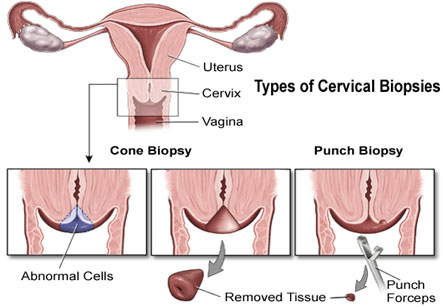
A cervical biopsy is a procedure which involves removal of a part of the cervix so the tissue can be examined under a microscope. The amount of cervical tissue removed depends on the method used:
• A simple cervical biopsy – It removes a small piece of tissue from the surface of the cervix.
• An endocervical biopsy – It removes tissue from high in the cervix by scraping with a scoop-shaped instrument called curette.
A cervical biopsy may be performed in a physician’s office on an outpatient basis. Procedures may vary depending on patient’s condition and the physician’s practices.
We have all the information you need about public and private clinics and hospitals that provide gynaecological treatment in Iran
Procedure:
• Patient is instructed to undress completely or from the waist down and put on a hospital gown.
• Bladder should be empty prior to the procedure.
• The position for the procedure is lying on the back on an examination table with feet raised and supported by foot rests.
• An instrument with smooth, curved blades known as speculum is inserted in the vagina. The speculum gently spreads apart the vaginal walls so the doctor can see inside the vagina and the cervix. The cervix is washed with a special solution and may be grasped and held in place with a clamp called a tenaculum.
• Often, the physician uses a colposcope, an instrument with a special lens similar to a microscope, for magnifying the cervical tissues. The colposcopeis placed at the opening of the vagina but does not enter the vagina.
• With the help of the colposcope, the doctor locates any problem areas on the cervix or in the vagina. Photographs with the colposcope or sketches of the areas on the cervix may be made for future record.
• The cervix may be cleansed and soaked with a vinegar solution, also called as an acetic acid solution. This solution stains the abnormal tissues in white and makes it more visible. A mild burning sensation may be felt during this process. An iodine solution may be used to coat the cervix, called as Schiller test.
• The type of biopsy performed is determined by the size, shape, location, and other characteristics of the abnormalities.
• The physician numbs the area using a small needle to inject anesthetic medication.
• For a simple cervical biopsy, one or more small samples of tissue are removed using a special type of forceps. A slight pinch or cramp may be felt while doing this. Cells from the inside of the cervical canal may be collected with a special instrument called an endocervical curette or an endocervical brush. This may also cause some cramping.
• For a cone biopsy, a larger cone-shaped piece of tissue is removed from the cervix. For this, the loop electrosurgical excision procedure (LEEP) or the cold knife cone biopsy procedure may be used. These procedures require the use of regional or general anesthesia.
• Bleeding from the biopsy site may be stopped with a paste-like topical medication. Electrocauterization (a probe with high frequency electrical signals to stop bleeding) or sutures may be required in some cases.
• After a cone biopsy, the cervix needs to be packed with a pressure dressing.
• The tissue is sent to a lab for examination.
After the biopsy:
Most women are able to resume normal activity the day of or within 1 day after the biopsy.
Some vaginal bleeding and a small amount of dark brown discharge are normal for about 1 to 2 weeks after biopsy.
Pads should be used instead of tampons for at least a week.
Sexual intercourse should be avoided for about 1 week.
Douching should not be done.
Indications:
A cervical biopsy is always be done before considering surgical treatment when abnormal tissue is seen on the vulva, vagina, or cervix, or if abnormal tissue is seen during colposcopy.
Results:
Results of the various tests such as abnormal Pap test, colposcopy, and cervical biopsy are compared and evaluated.
Results may indicate:
• Infection. The infection may be treated with antibiotics if it is caused by bacteria or yeast. Repeat Pap tests may be done to monitor the treatment.
• Minor cell changes. In these changes, only monitoring can be done without any treatment or treatment may be done to destroy or remove the abnormal cells. Over half of minor cell changes become normal on their own in 6 to 18 months.
• Moderate to severe cell changes. Treatment is required to destroy or remove the abnormal cervical cells.
• Cancer. Treatment is done to destroy or remove the tissue affected by invasive cancer.
• If the results of the initial abnormal Pap test, colposcopy, and cervical biopsy do not match, repeat Pap testing, with or without colposcopy, is recommended.
Risks
Vaginal bleeding can occur for up to 2 weeks after the cervical biopsy.